
Alzheimer’s may shrink the entorhinal cortex and amygdala up to a decade before memory loss
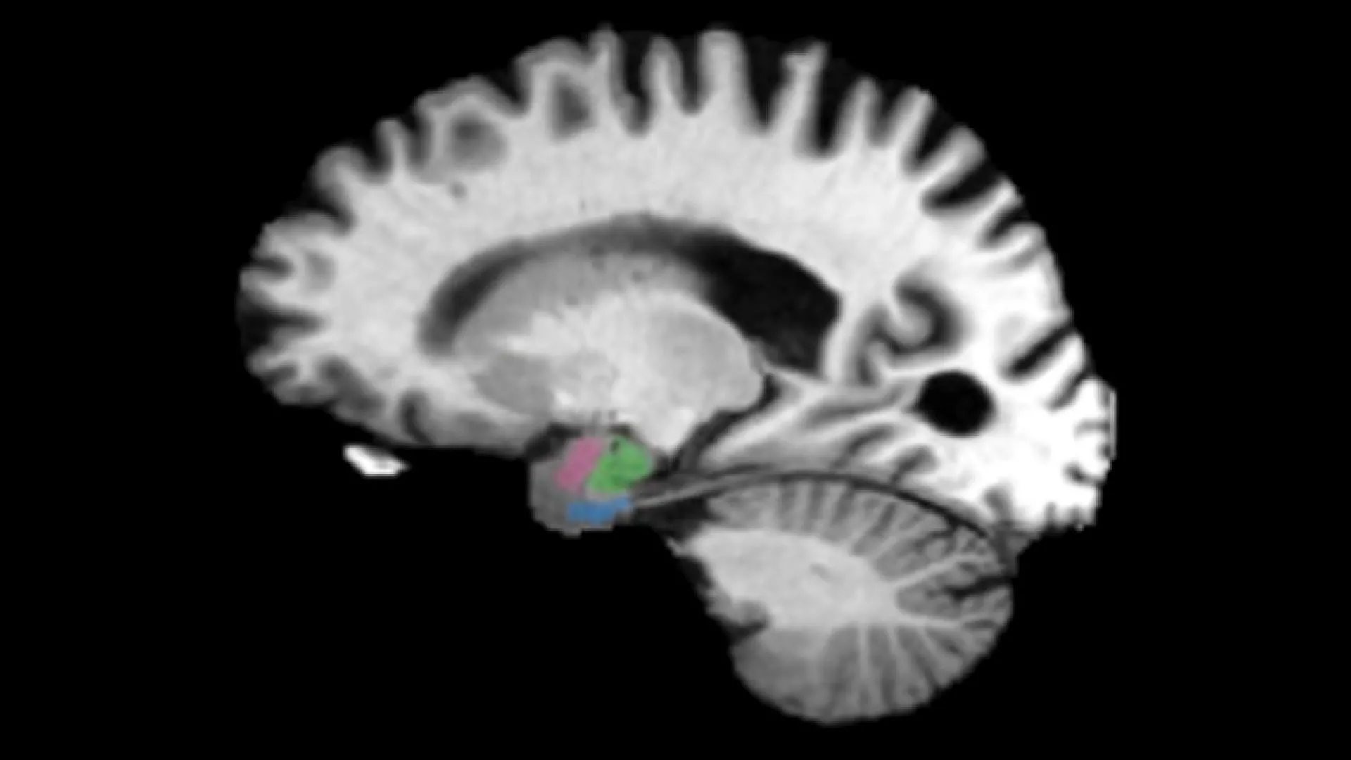
Johns Hopkins University researchers report that Alzheimer’s-related brain changes can be visible on scans as much as a decade before memory loss. In hundreds of MRI and PET scans from the Alzheimer’s Disease Neuroimaging Initiative and the Biomarkers of Cognitive Decline Among Normal Individuals study, the entorhinal cortex and amygdala—regions next to the hippocampus that help retrieve stored memories and process emotional memory—showed greater relative tissue loss in early stages than the hippocampus, the structure doctors usually watch. Participants ranged from people with no symptoms through cognitive impairment to patients who later died of the disease, and the pattern was consistent in both cohorts. The atrophy is tied to abnormal tau, a protein that normally supports nerve-cell structure. Senior author Michael Miller said clinical atrophy biomarkers appear at least five to ten years before symptoms, and that treating neurodegenerative change at two, three or four years would be far better than waiting until about fifteen years, when memory symptoms emerge. Co-author Marilyn Albert, director of the Johns Hopkins Alzheimer’s Disease Research Center, said the aim is to treat people before symptoms so decline might be delayed or stopped. Alzheimer’s is the leading cause of dementia worldwide and can progress silently for decades; the work, published in NeuroImage Reports and presented at Neuroscience 2026, the Society for Neuroscience’s annual meeting, points to checking these two regions together as an earlier detection path.
Continue in the app — vote & join in ➔